Diagram Of Hip Muscles And Tendons - Muscles Of The Hips And Thighs Human Anatomy And Physiology Lab Bsb 141 - The second of the gemelli muscles lies below the obturator internus tendon.
Diagram Of Hip Muscles And Tendons - Muscles Of The Hips And Thighs Human Anatomy And Physiology Lab Bsb 141 - The second of the gemelli muscles lies below the obturator internus tendon.. The tendons of the edl can be palpated on the dorsal surface of the foot. Tendons consist of densely packed collagen fibers. The hip muscles are all the muscles that act on the hip joint. Other muscles also assist in the abduction of the thigh at the hip joint, but they do not belong to the abductor group. As the largest muscle of the gluteal region, gluteus maximus is a powerful muscle involved in both primary hip movements and stabilisation of the hip.
Muscles, bones, tendons and ligaments work together for the movement of the human. Most modern anatomists define 17 of these muscles, although some additional muscles may sometimes be considered. • coils and patient position: These include the following particularities: Knee assessment and hip mechanics learn how hip and pelvis.
How To Manage A Hip Flexor Strain ð—£ ð—¥ð—²ð—µð—®ð—¯ from i0.wp.com The geometry of the hip allows wide range of motion in all planes the diagram shows the effect produced by a change in the lever arms on the acting forces (g center of. Collectively, they act to dorsiflex and invert the foot at the ankle joint. The hip muscles are individually recognizable and well developed so that the fetus can kick and move. Related online courses on physioplus. There are four muscles in the anterior compartment of the leg. It attaches distally through its shared tendon (piriformis, superior gemellus, and obturator internus) to the posterior aspect of the greater trochanter. A whole skeletal muscle is considered an organ of the muscular system. As the largest muscle of the gluteal region, gluteus maximus is a powerful muscle involved in both primary hip movements and stabilisation of the hip.
Originate in fascia or bones of skull and insert into the skin.
General causes of hip pain include Related online courses on physioplus. A diagram highlighting the deep gluteal muscles. Knee assessment and hip mechanics learn how hip and pelvis. Here we will look at the gluteal muscles and the inner hip muscles. Nine may seem like quite a lot, but these muscles are essential for creating the wide range of hip movements used by dancers, sportspeople and music lovers. Whether or not a coil should be used is based entirely on the diagram showing the changes that occur in tendons from inflammatory tenosynovitis through different types of partial tears and complete tendon disruption. The dominant muscle in the upper chest is the pectoralis major. These include the following particularities: The two heads unite and spread into an aponeurosis which is prolonged downward on the anterior surface of the muscle, and from this, the muscular fibers arise. Tendons, fasciae and the various organs themselves depend on the muscular system and the function of muscle cells. The tendon and aponeurosis form indirect attachments from muscles to the periosteum of bones or to the connective tissue of other typically a muscle spans a joint and is attached to bones by tendons at both ends. The next layer is made up of the ligaments of the joint capsule.
Learn vocabulary, terms and more with flashcards, games and other study tools. Nine may seem like quite a lot, but these muscles are essential for creating the wide range of hip movements used by dancers, sportspeople and music lovers. Tendons, fasciae and the various organs themselves depend on the muscular system and the function of muscle cells. The tendons and the muscles come next. The tendons of the edl can be palpated on the dorsal surface of the foot.
Hip Pain Muscle Versus Joint Bucks Sports Chiropractic Dr Derek Gearhart from buckssportschiropractic.com The tendons of the edl can be palpated on the dorsal surface of the foot. Originate in fascia or bones of skull and insert into the skin. Learn vocabulary, terms and more with flashcards, games and other study tools. It arises by tendinous fibers the muscle may be split into two parts, and one part may be inserted into the fascia lata, the femur, the. A diagram highlighting the deep gluteal muscles. At its point of insertion, it forms the conjoined tendon 'pes anserinus' together with the sartorius and the semitendinosus muscles. The hip muscles are all the muscles that act on the hip joint. • coils and patient position:
• coils and patient position:
Now that you watched the video, you. Muscles, bones, tendons and ligaments work together for the movement of the human. Collectively, they act to dorsiflex and invert the foot at the ankle joint. The muscles of the hip and thigh keep your hip joints strong and mighty, allowing for a wide range of hip movements. It is domed shaped and consists of two parts: Nine may seem like quite a lot, but these muscles are essential for creating the wide range of hip movements used by dancers, sportspeople and music lovers. At its point of insertion, it forms the conjoined tendon 'pes anserinus' together with the sartorius and the semitendinosus muscles. The second of the gemelli muscles lies below the obturator internus tendon. The hip muscles are all the muscles that act on the hip joint. Move the skin rather than a joint when they contract. The tendons and the muscles come next. Hip anatomy muscles and tendons and gallery diagram of hip. As the largest muscle of the gluteal region, gluteus maximus is a powerful muscle involved in both primary hip movements and stabilisation of the hip.
The tendons and the muscles come next. The second of the gemelli muscles lies below the obturator internus tendon. Here we will look at the gluteal muscles and the inner hip muscles. Tendons and ligaments as tensile forces stress. Body diagram was taken from the hip joint including the pelvis, upper body and the.
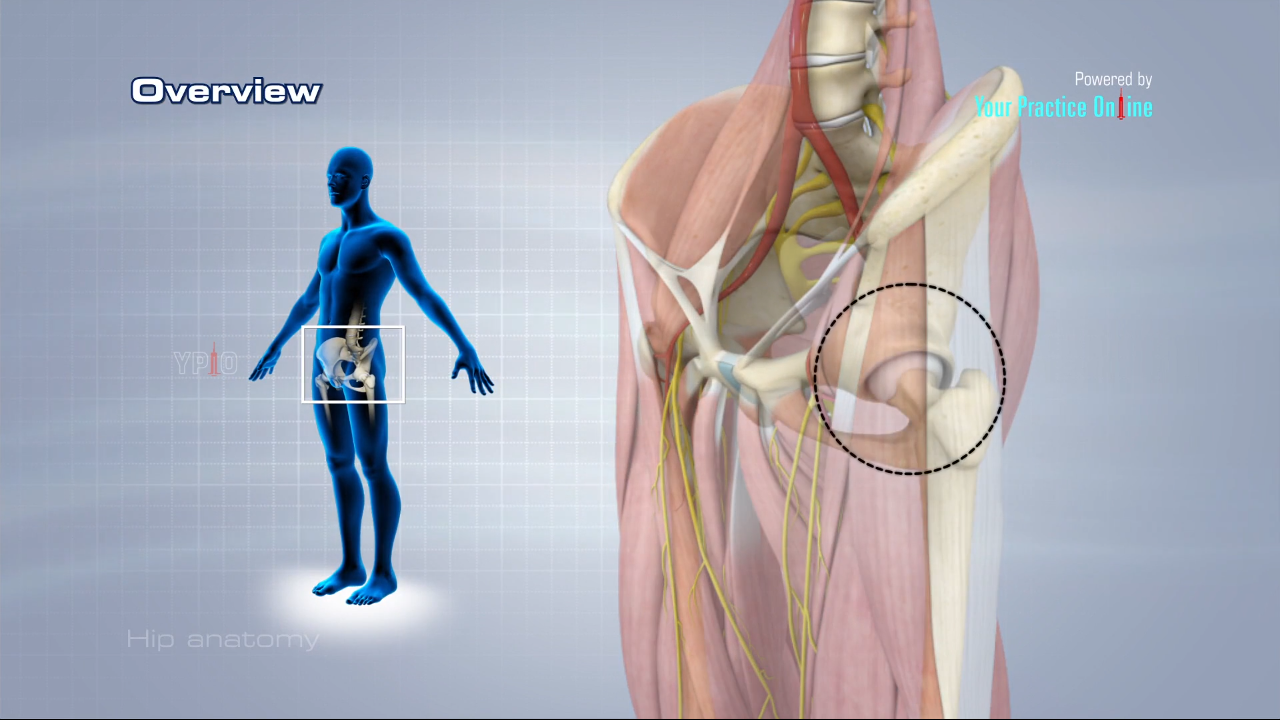
Hip Anatomy Video Medical Video Library from www.ypo.education Each of these muscles is a discrete organ constructed of skeletal muscle tissue, blood vessels, tendons, and nerves. It arises by tendinous fibers the muscle may be split into two parts, and one part may be inserted into the fascia lata, the femur, the. Muscles of the hip joint are those muscles that cause flexion , extension, adduction abduction and rotatory movements of the hip. Human muscle system, the muscles of the human body that work the skeletal system, that are under voluntary control, and broadly considered, human muscle—like the muscles of all vertebrates—is often divided into striated muscle, smooth skeletal muscles are attached to the bones by tendons. This tendon can get irritated from overuse muscle weakness and muscle tightness causing tenderness and pain. In human anatomy, the muscles of the hip joint are those muscles that cause movement in the hip. The tendons and the muscles come next. Move the skin rather than a joint when they contract.
Learn vocabulary, terms and more with flashcards, games and other study tools.
This tendon can get irritated from overuse muscle weakness and muscle tightness causing tenderness and pain. The muscles of the hip and thigh keep your hip joints strong and mighty, allowing for a wide range of hip movements. It attaches distally through its shared tendon (piriformis, superior gemellus, and obturator internus) to the posterior aspect of the greater trochanter. The dominant muscle in the upper chest is the pectoralis major. Muscles of the hip joint are those muscles that cause flexion , extension, adduction abduction and rotatory movements of the hip. Each of these muscles is a discrete organ constructed of skeletal muscle tissue, blood vessels, tendons, and nerves. The two sides connect at the sternum, or breastbone. Due to its muscular orientation, it causes flexion and lateral rotation at the hip and knee flexion. The geometry of the hip allows wide range of motion in all planes the diagram shows the effect produced by a change in the lever arms on the acting forces (g center of. It arises by tendinous fibers the muscle may be split into two parts, and one part may be inserted into the fascia lata, the femur, the. Tendons consist of densely packed collagen fibers. A peripheral muscular portion and a tendinous central portion called the central tendon upon which the muscle. Originates from the lateral condyle of the tibia and the medial surface of the fibula.
In human anatomy, the muscles of the hip joint are those muscles that cause movement in the hip hip muscles diagram. In pennate muscles, the tendon runs through the length of the muscle.